Tuesday, March 29, 2016

What Exactly Happens on the Cellular Level (With Fun Pictures!)

Pictured directly to the left is a cross-section of the the inside of a renal medulla (where filtration occurs). The small purple colored dots are the nuclei of the kidney’s epithelial cells which make up structures such as the walls of capillaries and microtubules. Because this is  cross section the actual tubule is white space surrounded by a ring of cells, like looking at a pipe through the hole. The orange arrow is pointing at an epithelial cell that has been altered by the cytopathic effects of the BK Virus. It becomes enlarged and as you can see the tubule will  begin to be closed up by the malformed cells, as well as the resulting scar-tissue.
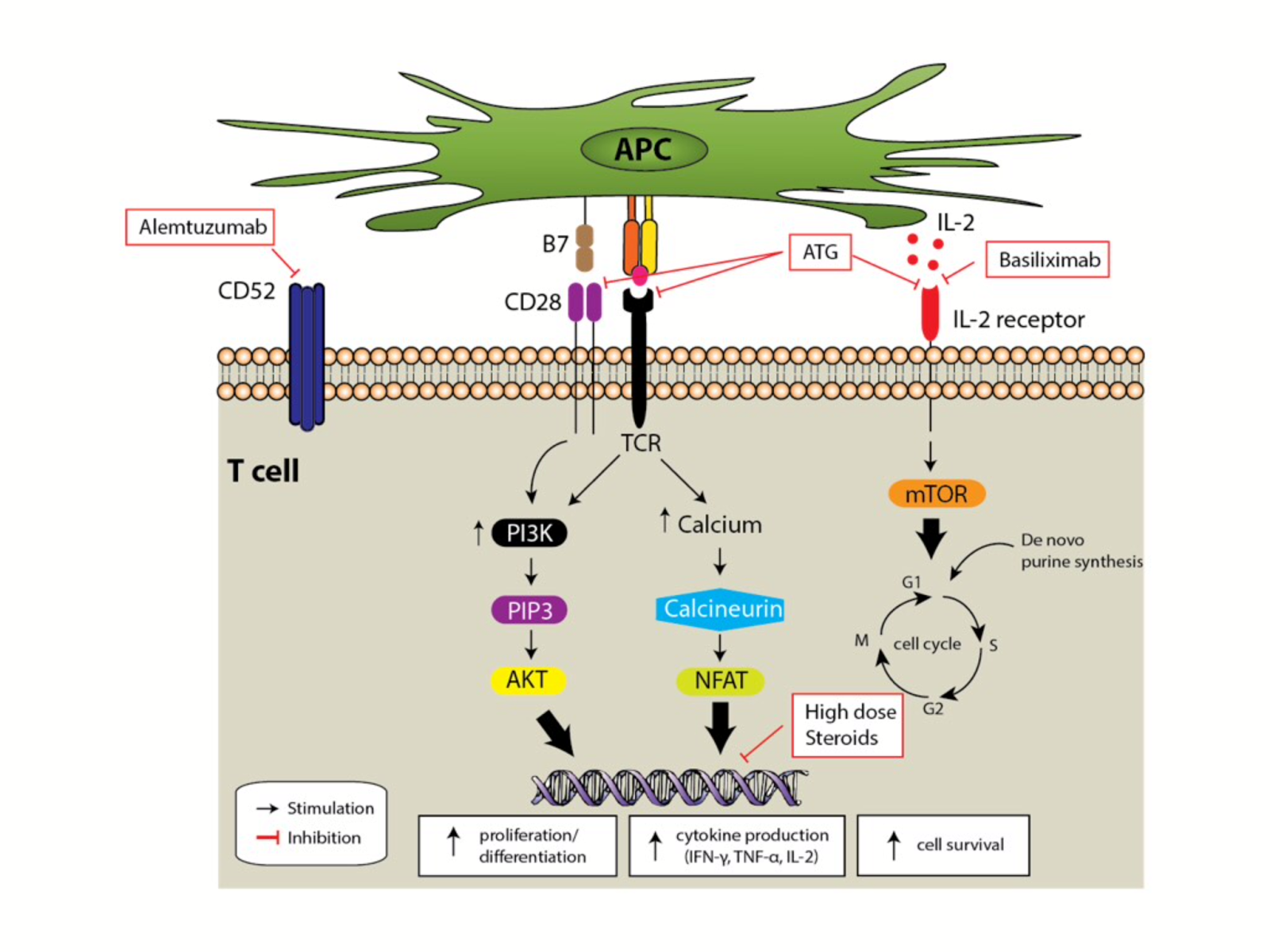
In the picture to the right, cells with a BK nuclear intrusion are highlighted using SV40 immunostaining. SV40 (Simian virus 40) is a virus in the same family as the BK (polyoma viruses) that can infect both humans and simians alike. Using it to stain locations of BK intrusion is another example of how we rely on research animals to be better able to help and understand ourselves, much like with rATG and hATG (see rATG post).
The final image shows fluorescent tagged immunoglobulin which is concentrated around areas of what is thought to be BK virus cytopathic manifestation.

BK Virus Nephropathy: History, Facts, and the Results of my Research on the Manifestations of the BK Virus (Part 2)

As mentioned in the previous post, it is for the most part unknown if a patient has progressed to nephropathy until the later stages of the disease. Because of this it is important to analyze a person’s risk for BKVAN by assessing them based on a number factors. Accurately predicting if someone is more likely to develop the disease preemptively is incredibly useful, as lowering the immunosuppressive load before onset is far more effective in BK Virus replication than doing so retroactively.
The risk factors can be broken down into three main categories: donor factors, transplant factors, and recipient factors. Donor factors are risk factors that relate to the condition of the person who is donating the kidney. These include BKV Seropositive status (whether the BK virus is in the blood of the donor) and absence of HLA C7 (important marker protein). The transplant factors mostly relate to the interactions between the donated kidney and the immune system of the recipient. These include whether or not (or to what extent) the kidney (or the surrounding tissues) was damaged during transplant, ABO incompatibility (differing blood types), and whether or not the recipient has had a rejection before. Typically if the transplant recipient has had previous transplants the immune system is already prepared to attack the graft and so rejection is more likely, but how this correlates to the recurrence of BKVAN is unknown. The final category, recipient factors, relate to various aspects of the host's body. They include things such as age, BK seronegative status, presence of diabetes, absence of HLA 7 and the need for immunosuppression.
After having compiled data for 237 HLA mismatched patients and 72 ABO-Incompatible patients between 1998 and 2010 I found a 6.1% and 17.8% prevalence of BKVAN respectively (p=0.08, a=0.05). Thus it can be seen beyond a reasonable doubt that the risk for developing it is severely higher when the graft and the recipient are not of the same blood type. While this correlation is important in and of itself, the reasons behind it are even more so. Because of the presence of such severely distinct markers on the graft, the recipient's immune system launches a much stronger attack on the new kidney thus necessitating a larger immunosuppressive load. The correlation between higher dosages of immunosuppressants and an increased risk for BKVAN gives credence to my initial hypotheses (see the first blogpost).

BK Virus Nephropathy: History, Facts, and the Results of my Research on the Manifestations of the BK Virus (Part 1)

BK Virus Nephropathy (BKVAN) is the clinical term for the condition in which the BK virus is actively harming the recipient's kidney graft. About 10% of renal transplant recipients will end up progressing from BK Viremia to BK Nephropathy. A plot of the data points of my collected data shows a slight upward increase in this number over the years 2000 to the 2014 ultimately leading to the most recent 10% statistic. A significant difference is found between the rates of incidence in first time and second transplant patients, with multi-transplant recipients being more at risk for nephropathy.
Though it is relatively easy to now detect whether or not the virus is in the bloodstream, it is much harder to see whether or not the viral infection has progressed to more severe stages. Renal biopsy (the sampling of some of the kidney tissue and then viewing it on a slide), remains the gold standard for recognizing BKVAN, however, many advancements in analyzing a hosts risk factors can lead to catching the disease in its earlier stages.
After a person has first come into contact with the virus (about 90% by the age of 23) it enters a state of non-replicative asymptomatic infection called “latency”. The very small virus particle embeds itself in the epithelial and urothelial cells in an immunocompetent host. Once an increased immunosuppressive load shifts the balance of immune system cells and virus particles towards the virus it begins to replicate and the graft goes through the three stages of BKVAN.
Stage A is where the virus starts to produce structural changes in the cells that make up the epithelial walls of the kidney. Unfortunately, it is hard for even a kidney biopsy conclusively determine that nephropathy is occurring at this early stage, often going undetected until it progresses to stage B. Stage B consists of  tubulointerstitial inflammation; the swelling of the capillaries used throughout the kidney to carry and filter blood. Stage C is tubular atrophy and interstitial fibrosis which severely reduces the kidneys functionality and fills it with scar tissue.
Between 30 to 60% of BKVAN cases progress to Stage C.

Sunday, March 27, 2016

What It Is and How It’s Made? The Synthesis of Rabbit ATG (rATG)

Used in over 55% of transplant cases rATG is by far the most commonly used. When most people are asked to imagine how it is synthesized most would say by a process of mixing solutions, enzymes, and proteins in a lab. This induction agent, however, is actually one two such agents made by immunizing another species to our white blood cells. Serums from over 26,000 (2012) immunized rabbits are pooled to ensure batch to batch consistency, an incredible effort on the part of both rabbits and humans.
The first report of immunizing an animal of one species against the immune cells of another species (mouse lymphocytes) was by Russian zoologist Ilya Metchnikoff. His trial (in 1899) involved injecting cells taken from mouse lymph nodes into a guinea pig. This guinea pig was then given time to develop antibodies specific to the immune cells of the mouse. A serum (ATG) was crafted from the blood of the guinea pig and then reinjected into other healthy mice. Metchnikoff observed a drop in the T cell count of the mice, and thus made an incredible breakthrough in the new field of immunology (and would later win a nobel prize for his work).
Our process for synthesizing rATG nowadays is still very similar to Metchnikoff’s. The notable difference is that instead of mice we immunize the rabbits to our own T cells. Rabbits are mainly utilized because of their ability to reproduce very quickly. The downside, that much like a blood donation for humans, they can only give so much within a given time. It is for this reason that sometimes equine ATG is also used, with a horse being able to give much more per donation. rATG and hATG are functionally identical and so they are chosen based on current price and availability.
It’s incredible to think that in order to keep one person’s organ alive and functional within another we appropriated the immune system response of another species.


Timeline for Important Immunosuppressants

As Mrs. Q has stated in a comment on an earlier post, the field of medicine has advanced rapidly in the past several decades and as new technologies and techniques were being discovered and perfected the margin for error has slowly decreased until it reached the accuracy we have today. The first truly successful kidney transplant didn’t occur until 1954 and was done between two identical twins, thus no immunosuppressive treatment was given or needed. Between 1954 and the present, many other kidney transplants were necessary, but early on, not many were attempted as the outcome would most surely be acute rejection unless the graft came from an identical twin (even then, mutations have a chance of affecting the expression of protein markers). It wasn’t until 1957, that the first widely used induction therapy agent Imuran, was used. With it came an incredible amount of side effects, the most severe being an increased chance of developing T cell cancer. From then until 1971, we did not know that the BK virus even existed, nor did we know of its detrimental effects on kidney grafts. Reliable detection would not come until 1983 and wouldn’t be perfected for another few years after this date. Because of this data from the before 1985 is very unreliable.
Another important factor which affects the reliability of the data was the immunosuppressive agents available and how widespread their usage was. It is for this reason that I have included at the end of this post a timeline with the year that important induction and maintenance agents were first synthesized, and for many when their usage became widespread.
eATG-- started in 1970, fully established by 1980 (Equine Anti-thymocyte Globulin)
rATG-- started 1980, fully established by 1990 (Rabbit Anti-thymocyte Globulin)
OKT 3-- 1985
Campath-- 2000
Basiliximab--1998
Daclizumab--1997
Imuran-- 1957
Celcept-- 1995
Cyclosporine-- 1983
Prograf-- 1994

Sunday, March 20, 2016

The Curious Case of the Regulatory T-Cell, Part 2

This T-Cell is another example of the body’s many self-check mechanisms which prevent excessive immune reactions, and stop an immune response once the invading organisms have been successfully eliminated. These cells are not naive T-Cells and therefore particular to only one antigen and work to stop NK cell and efferent T-Cells that are still attacking that antigen. This cell went unnoticed for a long time and is still hard to study today because it expresses nearly identical protein markers as the efferent T-Cells (the ones responsible for attacking antigens). Eventually after being isolated, it was noticed that an increase in the number of regulatory T-cells differentiated with respect to the markers on the graft found in the graft’s microenvironment leads to the formation of a protective barrier, offering localized antigen specific immunosuppression (the holy grail of transplantation). Another interesting relationship is how regulatory T-Cells interact with cancer. Cancer can be defined as a failure of the immune system to eliminate a cell which has suffered some kind of damage and now continues to replicate unchecked. One of the main reasons that the body will often not attack the tumor is because it expresses the same protein markers as other healthy cells in the body and so the body thinks that it is attacking itself. It is now thought that regulatory T-cells, in addition to stopping autoimmune disorders, also plays a critical role in protecting the cancer. It was noticed that if there was a high concentration of these types of cells in the cancer microenvironment it usually meant that the cancer prognosis was very poor. 
Research into these cells is still very new and a lot about how they function and how we can control their replication is still unknown. They may, however, prove to be very helpful in both finding a cure for cancer (or at least a way to diminish the effect of it) as well as opening the way for natural antigen specific immunosuppression.

The Curious Case of the Regulatory T-Cell, Part 1

Though BK nephropathy is a common risk of being on an immunosuppression regimen after transplantation (about 80% of the population is already infected with it) another major but less talked about risk is developing cancer. This is many times overlooked when one thinks of the negative effects of having an organ transplant, but the reality is that in order to keep a graft alive it is necessary to suppress the system in our bodies which fights cancer. However, good things can come from such morbid side effects as it is possible that we may have a found a potential way to both help increase the chance of graft survival and to reduce the malignancy of many cancers. Unfortunately, fighting cancer with this regimen may decrease the life expectancy of a graft, while supporting the graft may make it much harder to fight the cancer. The key to both treatments lies in how the elusive regulatory T-Cell functions.

Thursday, March 17, 2016

Immunosuppression, Maintenance Therapy

You probably could have guessed that this was coming after the last post. Even though induction therapy plays a large role in reducing the number of acute rejections, when trying to minimize the risk of long term rejection, maintenance therapy is key. Maintenance therapy is the immunosuppressant regimen that the patient will be on for the rest of their lives. A universal limitation on all immunosuppressive drugs is that they are non-antigen specific, and so because this regimen will affect the whole of the patient’s immune response a long period of time, patients must first be assessed based on their immunological risk before any kind of treatment can be started. Patients who have had previous transplants, are young, are African-American, or have an aggressive autoimmune disorder are at a higher risk for rejection and so are typically given higher dosages or more potent maintenance agents. The elderly, first transplant patients, and those without an autoimmune disease are generally low-risk. The immune system naturally breaking down with age is thought to be the reason older patients need less suppression, however, the flip-side of this is that older transplant patients have a longer recovery from the operation. Maintenance immunosuppression agents are usually used in conjunction with one another, especially when their effective mechanisms complement each other. The three main drugs are Calcineurin Inhibitors (CNI), mTOR Inhibitors and other anti-proliferative agents, and just like in induction therapy all of these are usually supplemented with steroids.
Because maintenance therapy is long-term suppression there are many side effects that arise from it, which generally do not arise from induction therapy (due to its very short duration). Above is a table I have found which shows just some of them for the varying drugs that are currently used.

Immunosuppression, Induction Therapy


Now that we have an understanding of how the immune system works, I will begin to discuss the main therapies used to prevent it from doing its job. Medicines used very soon after the surgery to suppress the initial immune response fall into the category of induction therapy. Those used to prevent rejection in the long term fall into the category of maintenance therapy. The goal of induction therapy is reduce the risk of acute rejection of the graft and as of 2015 80% of kidney transplant centers in the US use induction agents as part of their immunosuppression protocols. Induction agents are generally used because of their ability to affect several of the most critically important cells (T-Cells and B-Cells) and to quickly and severely reduce their effectiveness. It is for this reason that doctors will often put off a patient’s transplant even if they have a minor infection. This first wave of immunosuppressants can make even a small infection proliferate which may ultimately lead to death. The most commonly used agents are rATG (which is actually synthesized from rabbits!), Basiliximab, and Alemtuzumab. Each stops the naive T-Cell (T-Cell before it is presented with a particular antigen to attack) from becoming active in different ways, but one thing they all have in common is that they do not interfere with the patient’s recovery from the surgery. The most common maintenance agents (CNIs) have the side effect of constricting the blood vessels, which can prolong the amount of time before a wound (such as surgical cuts) heals. Alongside the induction agents transplant patients are given high-doses of steroids. The agents are generally taken only once, immediately after the procedure, however, the medicine has a long half-life (time it takes for it for half the medicine to be filtered out) and may stay in the body, working, for several weeks. Below is a picture of which part in T-Cell differentiation each medicine affects (I thought it looked cool).

Wednesday, March 16, 2016

General Immune System


The Two Categories
This post will be the first in a series of three describing what comprises the immune system as well as how various drugs and medical procedures, particularly those related to transplantation (more specifically the kidney), affect it. A human’s immune system can be broken down into two main categories, innate immunity and adaptive immunity. The main cells that form the innate response are NK cells, complement proteins, dendritic cells, and macrophages which act as a form of constant surveillance for any antigens. This form of response is characterized by its rapid action and the fact that it’s antigen nonspecific. It is “activated” by chemicals released by damaged tissues (cytokines) and generally forms the the initial strike against the transplanted organ. A surgery can very well be described as a “carefully coordinated attack on the body” and these attacks during transplantations lead to many ruptured vessels and separated tissues all of which “call for help”. Though the innate response cannot lead to the rejection of a graft itself, it plays a large role in augmenting the adaptive immune response. The adaptive immune response comprises mainly of T-Cells and B-Cells. B-Cells have the job of producing antibodies, which are Y-shaped proteins which function to identify particular antigens (each one is specific to one type of antigen). T-Cells further divide into Cytotoxic T-Cells and Helper T-Cells. Helper T cells are arguably the most important to mounting an immune response as they help dictate what antibodies the B-Cells produce and also activate Cytotoxic cells to destroy the antigen. Though the adaptive response is much slower than the innate response, this is the one that ultimately leads to the destruction of the graft. Though it takes time for the correct antibodies to proliferate and for the T-Cells to target the kidney, it is a very specific attack on only those foreign cells, and it will continue until the failure of the graft (unlike the innate response which dwindles as the cytokines do). It is the careful and balanced suppression of this response that nephrologists (as well as cardiologists and gastroenterologists) seek for the survival of the graft.

Wednesday, March 9, 2016


Are things getting worse or are we just now discovering how bad they were? One of the most difficult parts of analyzing apparent trends in the incidence of BK viremia is attributing it to the proper source. I in particular am now looking at the specific times after which new immunosuppressive agents began to be used more regularly and seeing if the graphed incidence rates I have compiled thus far show any significant changes at or around those times. One thing that may, however, be a confounding variable for the overall trend is the many advances we have made in virus detection over the past three decades. This could potentially mean that many graft losses in the past may have misattributed to causes other than BK viremia simply due to the uncertainty then found in detection techniques. The main way to detect polyomaviruses in general is the Urinary EM, to check for the BK virus specifically, however, Polymerase Chain Reactions are used. This is a not a new process (being first done successfully in 1983), but like with most things we’ve polished and and refined it, slowly eliminating as much room for error as possible. Due to the time it took to figure out the optimal incubator times, temperatures and which ions disrupt the process (Mn2+) initial results had a greater risk for error than those we get now.  As a result, I have one more thing to account for when to research this week in order to ensure that my data is meaningful. (Image is of 1 BK Virus)